3D anatomy atlas
Learn anatomy on 3D models, in your language
24 atlases built from open data: whole bodies, regions, organs and 12 real CT cases. Rotate, cut, pull apart and test yourself, right in the browser.

3D models: BodyParts3D, © The Database Center for Life Science licensed under CC Attribution 4.0 International
Atlases
See all atlases (24)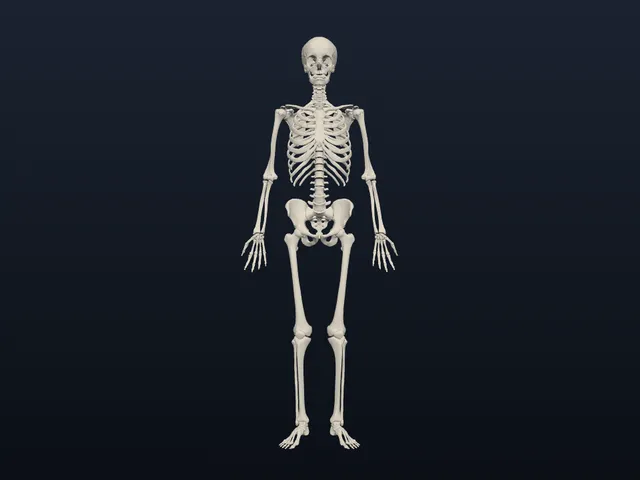
Adult male (BodyParts3D)
Whole bodyCC BY 4.03,432 structures
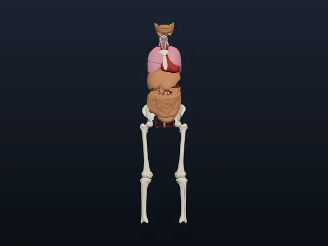
Adult female organs (HuBMAP HRA)
Whole bodyCC BY 4.01,166 structures
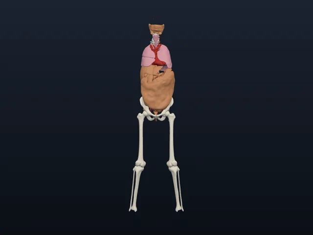
Adult male organs (HuBMAP HRA)
Whole bodyCC BY 4.01,132 structures
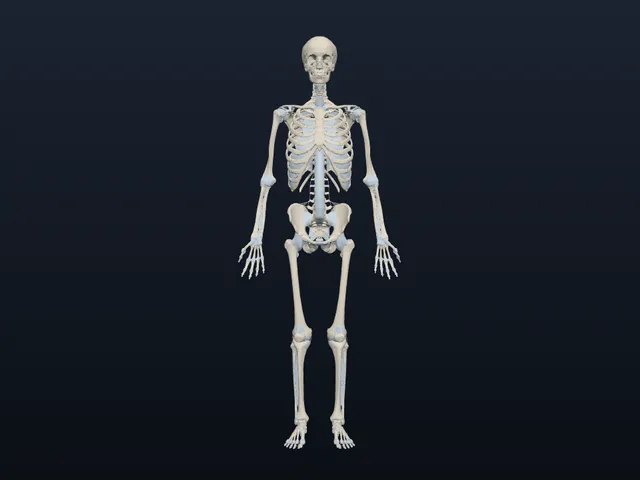
Adult male, detailed (Z-Anatomy)
Whole bodyCC BY SA 4.0Share-alike5,461 structures
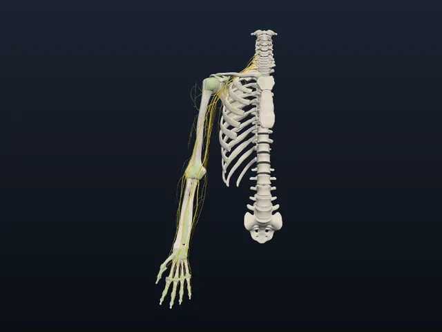
Right upper limb (Open3DModel)
RegionCC BY SA 4.0Share-alike707 structures
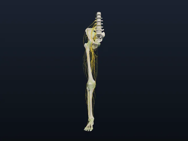
Right lower limb (Open3DModel)
RegionCC BY SA 4.0Share-alike613 structures
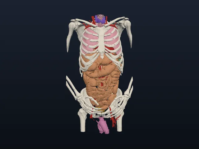
Male torso and inner organs (Voxel-Man, Visible Human)
RegionCC BY 4.0232 structures
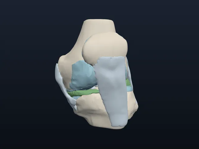
Knee (SPL atlas)
Region3D Slicer B48 structures
Start learning
All guidesAnatomical planes and sections
This page explains sagittal, coronal and transverse anatomical planes, what their sections show, and how they relate to CT, MRI and oblique slices.
Anatomical position and directional terms
This page explains anatomical position, paired directional terms, and how to describe relationships between structures without confusing left and right.
Anatomical terminology: Latin names, TA2 and FMA
This page explains Latin anatomical names, Terminologia Anatomica, TA2 numbers, FMA and UBERON identifiers, and how to interpret names on structure pages.
Anatomical variation: why bodies differ from the atlas
Anatomical variation explains how bodies differ in structure, why textbook patterns are not universal, and how sex, age and real-case atlases add context.
Arteries, veins and the two circulations
Learn how systemic and pulmonary circulation connect, how artery and vein walls differ, and how great vessels, portal vessels and anastomoses fit.
Body cavities and serous membranes
This page explains the major body cavities, their contents, the diaphragm’s role, and the parietal and visceral layers of serous membranes.
Study tools
-
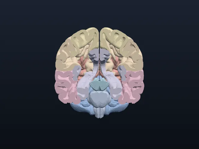
Cross-section
Cut the model along three planes; cut faces are filled layer by layer.
-
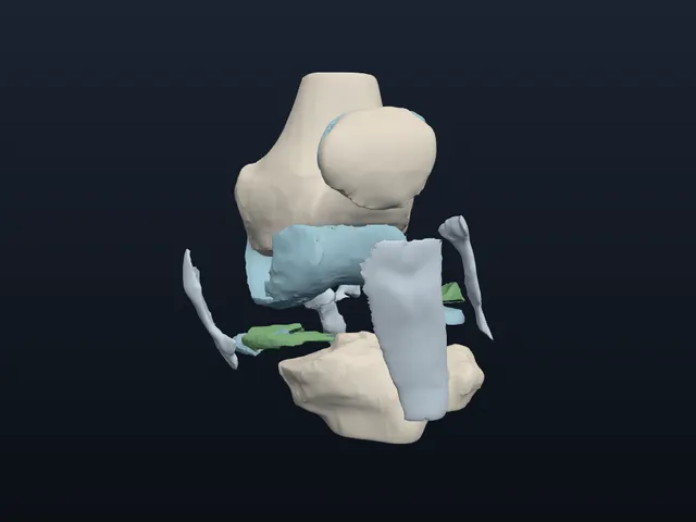
Explode
Pull the parts apart to see how they fit together.
-
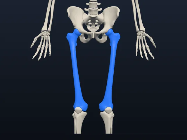
Self-test
Find and name structures on the model, with spaced repetition.